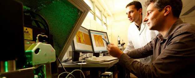
We
have our own
facilities at McGill University, which include nonstandard experimental
setup for unique experiments at small length scales and
in-situ
(we image the material at high and low magnifications in
real time as the material is deformed or fractured).
Miniature Loading Stage
| We use this small
mechanical loading stage for a wide variety of tests, from cyclic
loading on small mice bones to fracture tests on seashells. The stage
is small enough to fit under our optical microscope or our scanning
probe microscope. This allows for in-situ testing, where we can image
the material as it is deformed. The stage is controlled with a
computer-controlled (with feedback on force or displacement) which
enables a wide variety of testing programs. We use this machine with
different types fixtures, some custom made. These include tensile, 3
and 4 point bending, compression, indentation. Our load cells include
100 lbs, 25 lbs,5 lbs and 20 grams |

|
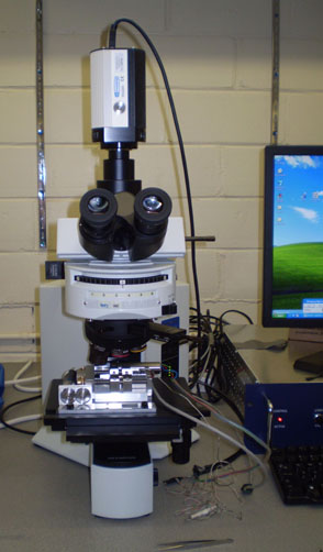
Olympus upright optical
microscope
We use this microscope for a variety of purposes, mostly microstructure
characterization for which we have differential interference contrast
and white light / fluorescence capabilities and a high resolution CCD
camera. Our mechanical loading stage also fits on the microscope stage
so we can image samples as they are deformed. An important implication
is that we can use the images to measure strain with a great accuracy,
using digital image correlation.
|
|
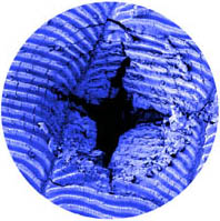
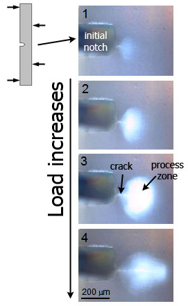
Example of in-situ
imaging of a propagating crack in nacre from a seashell
|
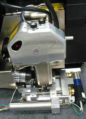
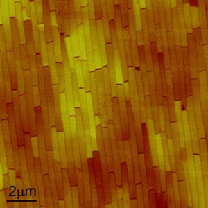
Veeco Nanoman Scanning Probe Microscope (SPM)
The
scanning
probe microscope is an extremely versatile piece of equipment used for
imaging surfaces with nanometers resolution but also to actually
interrogate surface mechanically (elasticity, strength, friction...) ,
or even pull on single molecules (force spectroscopy). An interesting
setup is to fit the mechanical stage underneath the microscope in order
to monitor how the microstructure of materials evolves when mechanical
stresses are applied. This setup allows great image resolution on
sample in wet conditions, making it perfect for in-situ mechanical
tests on hydrated biological materials.
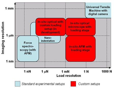
Intermediate load
mechanical testing
platform (in construction)
Large
specimens and
structures are tested on a traditional universal tensile machine, while
on the other end the mechanics of single molecules can be interrogated
using an atomic force microscope (AFM). Between these two extremes
there is a wide gap in terms of force and imaging resolution, which we
are filling with new :intermediate" experimental setup.
We also use the
following equipements
and facilities:
*
Nanoindenter (Hysitron Triboscope) used to "poke" materials at depth of
micrometers down to a few nanometers
* Universal tensile machine (UTM) used
to test large
samples or structures
* Precision diamond saw used to carve
millimeter
size samples for mechanical testing
* Polishing facilities to prepare
surfaces for
imaging
* Electron Microscopes at the
FEMR
* Micro Computer Tomography at
the
Bone
center
* Chemical Facilities in the Otto Mass
building
* Microfabrication at the
McGill
Nanotools microfab (MNM)